
Anatomical variations of caecal appendix on tomography, a retrospective study.
Variaciones anatómicas del apéndice cecal en tomografía, un estudio retrospectivo.
Barra lateral del artículo
 FLIP
FLIP

Contenido principal del artículo
Resumen
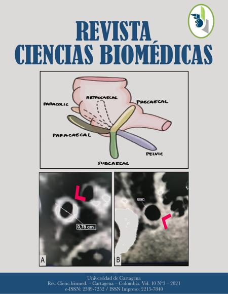
Introduction: the most common surgical cause of abdominal pain is appendicitis; its diagnosis is affected by anatomical variations of the vermiform appendix, because this is the most variable abdominal organ in terms of position and organ relations. Objective: to determine the characteristics of the normal appendix in computed tomography scans, including length, diameter, wall thickness, and the location of the base and the appendicular tip. Methods: abdominal computed tomography scans with UroCT scan protocol images were studied to measure length, width, and diameter of the appendix, and to identify the locations of its base and tip. The appendicular tip location was categorized as anterior or posterior and subdivided into: pelvic, retrocaecal/retrocolic, postileal, paracolic, subcaecal, subhepatic or midline. The appendicular base location was defined in three planes in relation to the ileocaecal valve: anterior-posterior, medial-lateral, or superior-inferior. Results: were included the abdominal tomography images of 83 patients in which general characteristics of the appendix were determined: a mean length of 78 mm (SD = ±23.41) and a mean diameter of 6.55 mm (SD =± 1.77) were observed. The bases were more frequently located inferior posterior, lateral and medial, to the ileocaecal valve, and the tips were more frequently located in the pelvic cavity, followed by the retrocaecal and paracaecal location. Conclusions: the locations of the base of the appendix were correlated with overall reports. There was no correlation between the tip location and the length of the appendix, this means that, even if the appendix is long, it is not associated with a tip location further from the ileocaecal valve.
Palabras clave
Descargas
Datos de publicación
Perfil evaluadores/as N/D
Declaraciones de autoría
- Editor y equipo editorial
- Perfiles
- Sociedad académica
- Universidad de Cartagena
- Editorial
- Universidad de Cartagena

Para obtener más información sobre un dato, haga clic ![]()
Public Facts Label - Plugin Mantenido por Public Knowledge Project
Detalles del artículo
Referencias (VER)
Snyder MJ, Guthrie M, Cagle S. Acute appendicitis: Efficient diagnosis and management. Am Fam Physician. 2018; 98(1): 25–33.
Cabrera-Rivera PA, Posso-Valencia HJ, Dennis-Verano RJ. Clinical and cost benefits of a standardization model in the management of acute appendicitis. Rev Colomb Cir. 2021; 36(2): 283–300. https://doi.org/10.30944/20117582.630 DOI: https://doi.org/10.30944/20117582.630
Monsonis B, Mandoul C, Millet I, Taourel P. Imaging of appendicitis: Tips and tricks. Eur J Radiol. 2020; 130: 109165. Available from: https://doi.org/10.1016/j.ejrad.2020.109165 DOI: https://doi.org/10.1016/j.ejrad.2020.109165
Banerjee A, Kumar IA, Tapadar A, Pranay M. Morphological Variations in the Anatomy of Caecum and Appendix - A Cadaveric Study. Natl J Clin Anat. 2012 Jan 23; 01(01): 030–5. Available from: http://www.thieme-connect.de/DOI/DOI?10.1055/s-0039-3401654 DOI: https://doi.org/10.1055/s-0039-3401654
Zacharzewska-Gondek A, Szczurowska A, Guziński M, Sąsiadek M, Bladowska J. A pictorial essay of the most atypical variants of the vermiform appendix position in computed tomography with their possible clinical implications. Polish J Radiol. 2019; 84: e1–8. https://doi.org/10.5114/pjr.2018.81158 DOI: https://doi.org/10.5114/pjr.2018.81158
Ghorbani A, Forouzesh M, Kazemifar AM. Variation in Anatomical Position of V ermiform Appendix among Iranian Population: An Old Issue Which Has Not Lost Its Importance. Anat Res Int. 2014 Sep 10; 2014: 1–4. Available from: https://www.hindawi.com/journals/ari/2014/313575/ ; https://doi.org/10.1155/2014/313575 DOI: https://doi.org/10.1155/2014/313575
JM W, W M, JL C. Does this patient have appendicitis? JAMA. 1996; 276(19): 1589–94. https://doi.org/10.1001/jama.1996.03540190061030 DOI: https://doi.org/10.1001/jama.276.19.1589
Johnson PT, Eng J, Moore CJ, Horton KM, Fishman EK. Multidetector-row CT of the appendix in healthy adults. Emerg Radiol. 2006; 12(6): 248–53. https://doi.org/10.1007/s10140-006-0491-y DOI: https://doi.org/10.1007/s10140-006-0491-y
Mwachaka P, El-busaidy H, Sinkeet S, Ogeng’o J. Variations in the Position and Length of the Vermiform Appendix in a Black Kenyan Population. ISRN Anat. 2014 Apr 30; 2014: 1–5. Available from: https://www.hindawi.com/journals/isrn/2014/871048/ ; https://doi.org/10.1155/2014/871048 DOI: https://doi.org/10.1155/2014/871048
Charoensak A, Pongpornsup S, Suthikeeree W. Wall thickness and outer diameter of the normal appendix in adults using 64 slices multidetector CT. J Med Assoc Thail. 2010; 93(12): 1437–42.
Baird DLH, Simillis C, Kontovounisios C, Rasheed S, Tekkis PP. Acute appendicitis. BMJ. 2017; 357: j1703. https://doi.org/10.1136/bmj.j1703 DOI: https://doi.org/10.1136/bmj.j1703
Kabir SA, Kabir SI, Sun R, Jafferbhoy S, Karim A. How to diagnose an acutely inflamed appendix; a systematic review of the latest evidence. Int J Surg. 2017; 40: 155– 62. Available from: https://doi.org/10.1016/j.ijsu.2017.03.013 DOI: https://doi.org/10.1016/j.ijsu.2017.03.013
Demir MK, Savas Y, Furuncuoglu Y, Cevher T, Demiral S, Tabandeh B, et al. Imaging Findings of the Unusual Presentations, Associations and Clinical Mimics of Acute Appendicitis. Eurasian J Med [Internet]. 2017 Oct 25; 49(3): 198–203. Available from: https://www.eajm.org//en/imaging-findings-of-the-unusual-presentations-associations-and-clinical-mimics-of-acute-appendicitis-132995 ; https://doi.org/10.5152/eurasianjmed.2017.17218 DOI: https://doi.org/10.5152/eurasianjmed.2017.17218
Whitley S, Sookur P, McLean A, Power N. The Appendix on CT. Clin Radiol. 2009; 64(2): 190–9. https://doi.org/10.1016/j.crad.2008.06.015 DOI: https://doi.org/10.1016/j.crad.2008.06.015
Pinto P, N PL, Jm P, Cunha R, Pinto P. CT Evaluation of Appendicitis and Its Complications : Am Roentgen Ray Soc. 2005; 185 (August): 406–17. https://doi.org/10.2214/ajr.185.2.01850406 DOI: https://doi.org/10.2214/ajr.185.2.01850406
Giovanni E, Gómez C, Luis P, Porras F, Uribe LA, Camargo DB. Posición anatómica y longitud del apéndice vermiforme en una población de raza mestiza de la ciudad de Bucaramanga - Colombia. 2009; 55: 116–20.
Barlow A, Muhleman M, Gielecki J, Matusz P, Tubbs RS, Loukas M. The vermiform appendix: A review. Clin Anat. 2013 May; 842. Available from: https://onlinelibrary.wiley.com/doi/10.1002/ca.22269 ; https://doi.org/10.1002/ca.22269 DOI: https://doi.org/10.1002/ca.22269
Willekens I, Peeters E, De Maeseneer M, de Mey J. The Normal Appendix on CT: Does Size Matter? Lo AW, editor. PLoS One. 2014 May 6; 9(5): e96476. Available from: https://dx.plos.org/10.1371/journal.pone.0096476 ; https://doi.org/10.1371/journal.pone.0096476 DOI: https://doi.org/10.1371/journal.pone.0096476
Wakeley CPG. The Position of the Vermiform Appendix as Ascertained by an Analysis of 10,000 Cases. J Anat. 1933; 67(Pt 2): 277–83.


